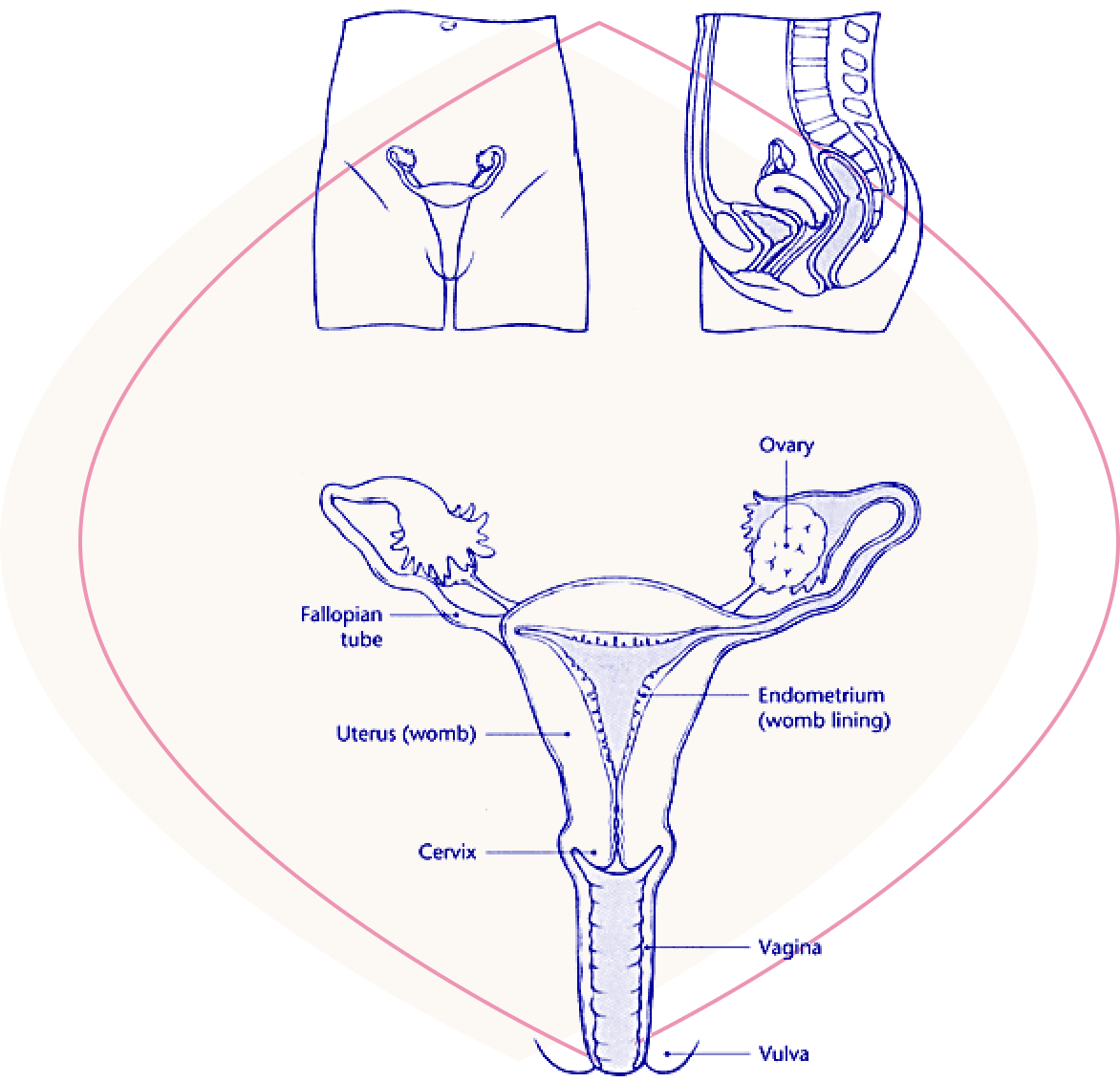
The female genital tract includes the vulva, vagina, cervix,
uterus, Fallopian tubes and ovaries.
The vulva is the fleshy folds that surround the opening to the
vagina.
The vagina is a muscular canal which lies between the vulva and
the cervix.
The cervix is a 'barrel-shaped' organ which lies at the end of
the vagina. It is sometimes called the 'neck of the womb'.
The uterus (or womb) extends from the cervix and lies within the
pelvis. The uterus is the organ in which a baby develops.
The endometrium lines the uterus. This lining becomes
progressively thicker towards the middle of the menstrual cycle
in preparation for the fertilized egg. If fertilization does not
occur, the top layer of the endometrium breaks down and is lost
during menstruation.
The Fallopian tubes connect the uterus to the ovaries. When an
egg is released by an ovary, it passes into the Fallopian tube,
where it is fertilized, and then passes down into the uterus.
The ovaries lie at the end of each Fallopian tube. Every month,
an egg is produced from one of the ovaries and develops in a
small 'cyst' (or follicle).
In 75% of women, the uterus lies forwards towards the pubic bone
and is said to be 'anteverted'. In the remaining 25% of women,
the uterus lies backwards and is said to be 'retroverted'.
Certain conditions, such as endometriosis, can cause a uterus
that is normally anteverted to become retroverted, which may
cause pelvic pain. Very occasionally, an operation may be
necessary to correct this.
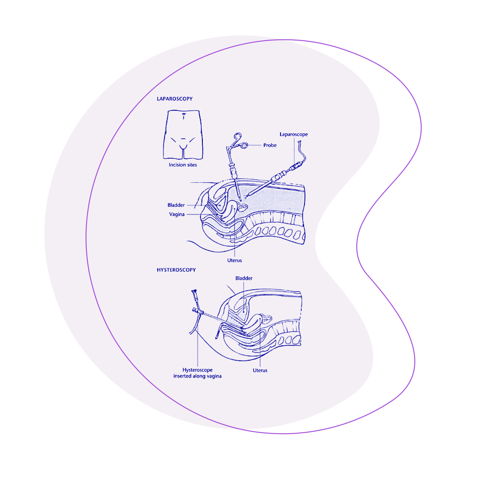
Laparoscopy is used to examine the abdomen to investigate
pelvic pain, ectopic pregnancy and
infertility. Hysteroscopy is used to examine the uterus
to investigate heavy and/or irregular menstrual bleeding,
or postmenopausal bleeding. In both procedures, a
small, fibre-optic 'telescope' is used to see the internal
organs.
Both laparoscopy and hysteroscopy are usually performed
under a general anaesthetic as a day-case procedure and
take 15-20 minutes. Hysteroscopy can also be performed
under local anaesthetic, as an outpatient procedure.
In laparoscopy, the instrument is passed through a
small incision in the abdomen. A second incision may
be made so that a probe can be inserted to manipulate
the organs. Carbon dioxide gas is then pumped into
the abdomen to separate the tissues so that the organs
can be seen more clearly.
In a hysteroscopy, the instrument is passed along
the vagina and through the cervix in order to examine
the lining of the uterus. No incision is made.
Patients usually recover from hysteroscopy
rapidly. Following laparoscopy, however, patients
may experience a pain similar to a period pain or bloating
for up to 1 week.
Minimally invasive or 'key-hole' surgery can be
performed using laparoscopy and hysteroscopy. This has
the advantages of reduced postoperative pain, a
shorter hospital stay, a smaller incision and therefore a
better cosmetic results than conventional surgery.
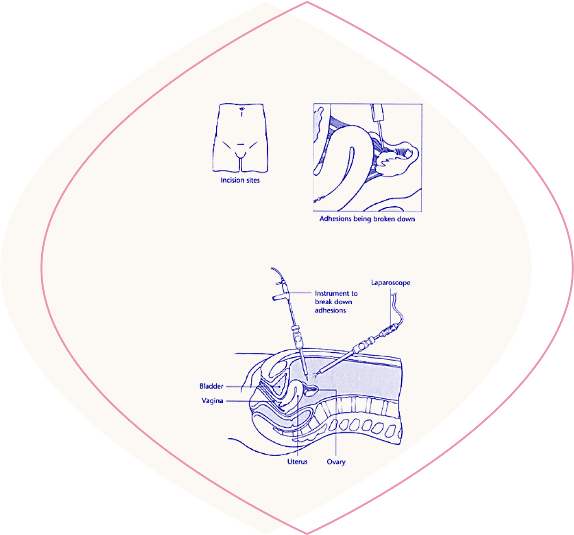
Laparoscopic adhesiolysis is used to remove scar
tissue called adhesions, which are caused by infection
or previous surgery, in patients with chronic pelvic pain
or infertility. As a result of this scar tissue, organs
which are normally separate, such as the bladder and bowel,
can become 'stuck' together causing pain.
The operation is performed under a general anaesthetic, and
the length of time taken will vary from 30 minutes to
2 hours depending on the number of adhesions.
The procedure is performed laparoscopically, using a small,
fibre-optic 'telescope', which is inserted into the abdomen
through a small incision. The adhesions are broken down
with specially designed instruments. All the organs remain
intact.
There will be some discomfort following surgery which
is controlled with pain killers.
The average hospital stay is overnight and
complete recovery should occur within one week.
Dilatation and curettage (D & C or uterine evacuation)
Dilatation and curettage (D & C) may be necessary
to investigate abnormal, heavy or irregular bleeding, or
to remove what are called the 'products of
conception' following miscarriage.
Nowadays, a hysteroscopy to examine the lining of
the uterus is often carried out before a D & C. This
involves passing a small, fibre-optic 'telescope' along the
vagina and through the cervix in order to examine the
lining of the uterus.
D & C is usually performed under a general
anaesthetic as a day-case procedure and takes about 15
minutes.
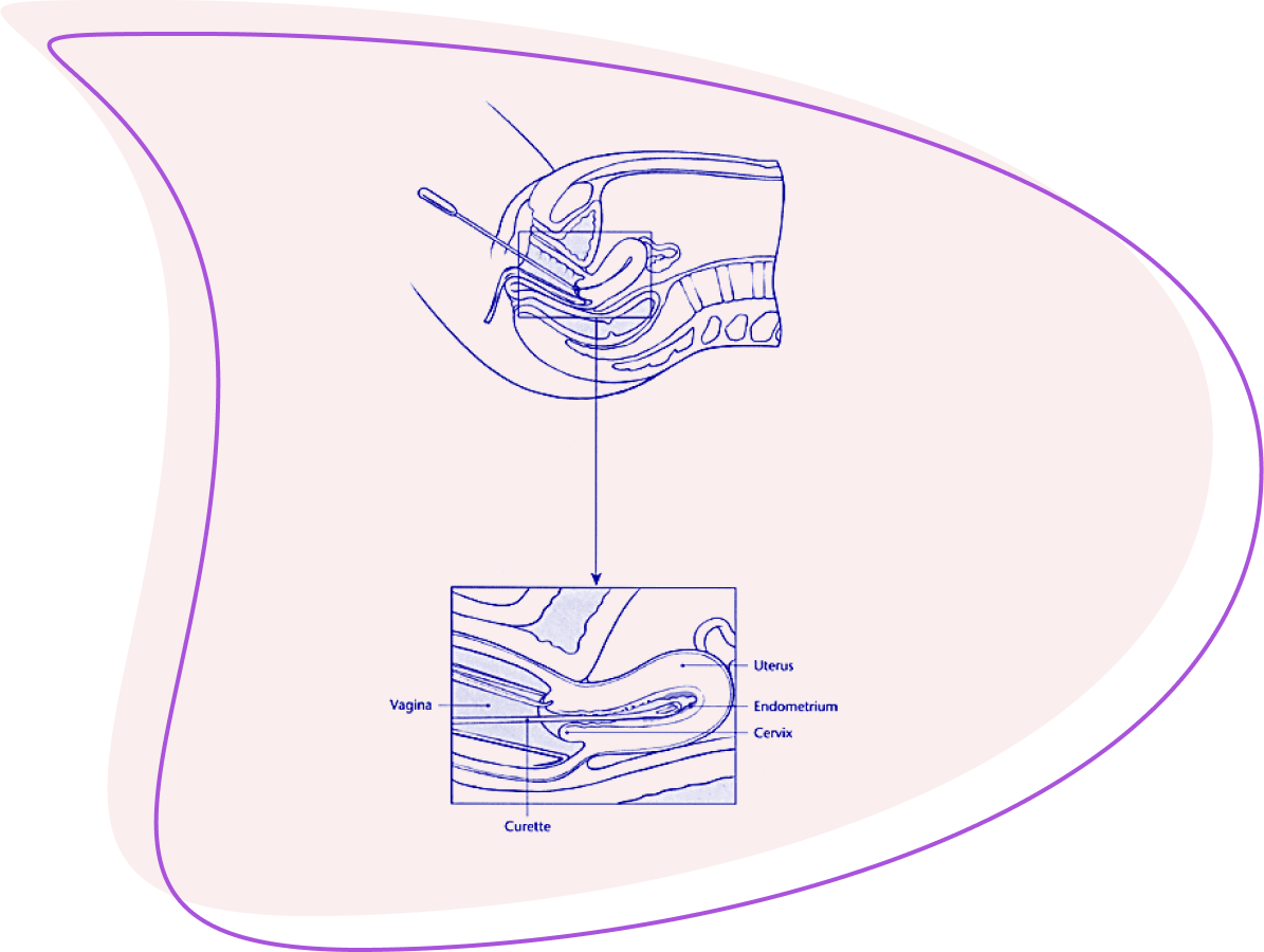
The cervix is dilated with a smooth instrument called
a dilator. A curette, which is a small, spoon-shaped
instrument, is then passed through the cervix to remove the
tissue lining the uterus (the endometrium). The tissue is
then sent to the laboratory to be examined.
Patients usually recover rapidly, but may
experience discomfort similar to a period pain for a few
hours after the procedure. There may also be a small amount
of bleeding, which may last 1-2 days.
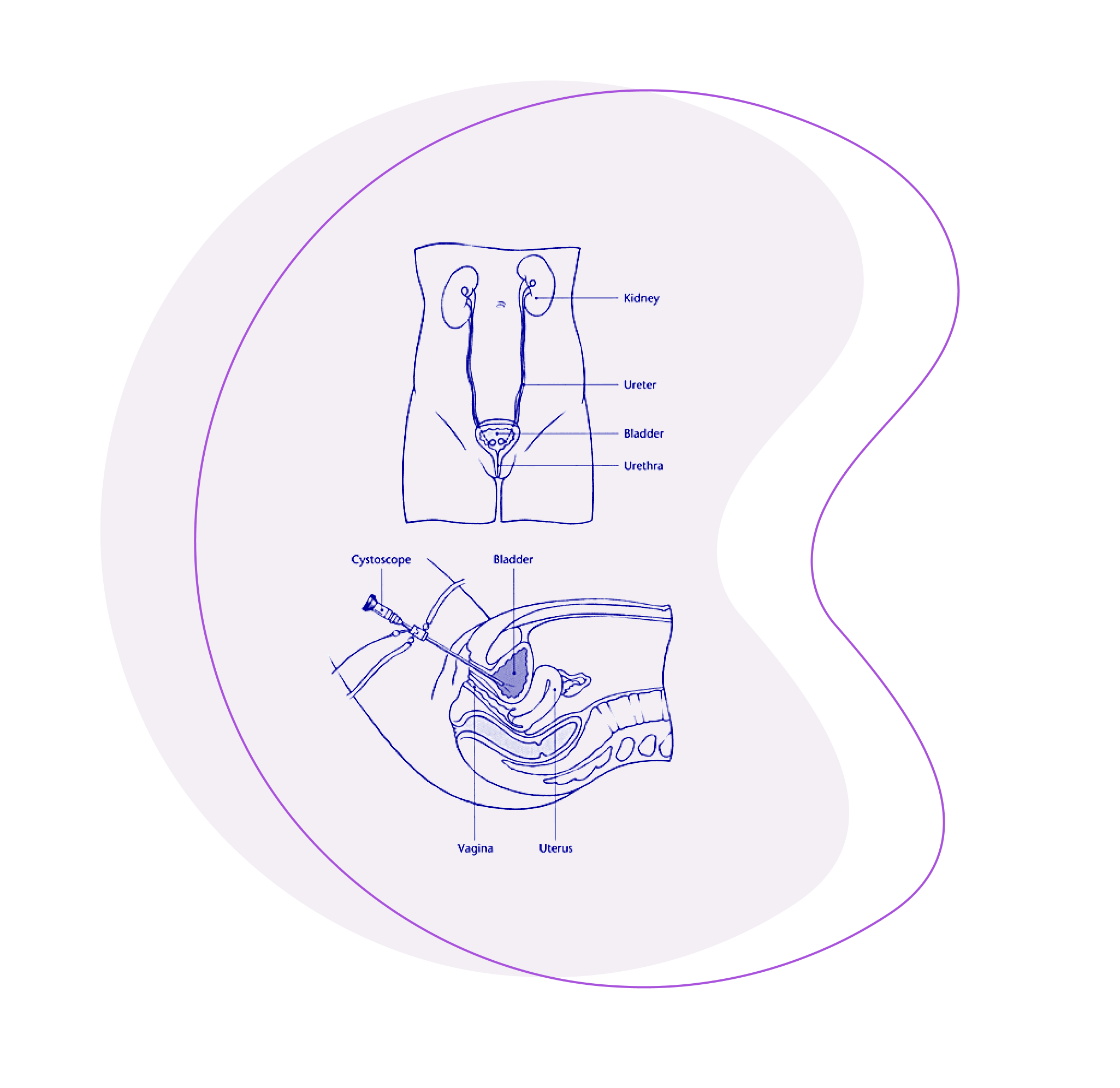
Cystoscopy is examination of the lining of the
bladder using a thin, fibre-optic 'telescope'.
The procedure is used to investigate the cause of urinary
symptoms, such as urgency, frequency or blood in the urine,
or as part of the investigation of cancer of the cervix.
Cystoscopy is usually performed under a general anaesthetic
as a day-case procedure and takes about 5-10 minutes.
The urethra (the tube through which urine passes from the
bladder to the outside) is dilated and the bladder filled
with fluid through a soft plastic tube called a catheter.
The cystoscope is then inserted to examine the lining of
the bladder.
Normal activities can be resumed the following day.
In patients undergoing major abdominal surgery (e.g.
for cancer of the cervix or ovary), cystoscopy may be used
to insert plastic stents into the ureters (the tubes
which drain the urine from the kidneys into the bladder),
so that the surgeon can identify them more easily. The
stents are removed after surgery.